X-ray exposure factors chart pdf
OPERATOR’S INSTRUCTIONS This X-ray equipment may be dangerous to patients and operators unless safe exposure factors and operating instructions are observed.
Exposure adjustment systems can be categorised into simple techniques, which minimises the exposure adaptation steps, mathematical approaches, comprehensive charts and technological approaches which eliminate mental calculations and chart searching through the use of technology.
10 standardized radiation exposure, KSTD, that would produce the same detector response. 11 The regions where the median is determined may be defined in different ways (Section 5 12 Assessment of Detector Response, K IND ).
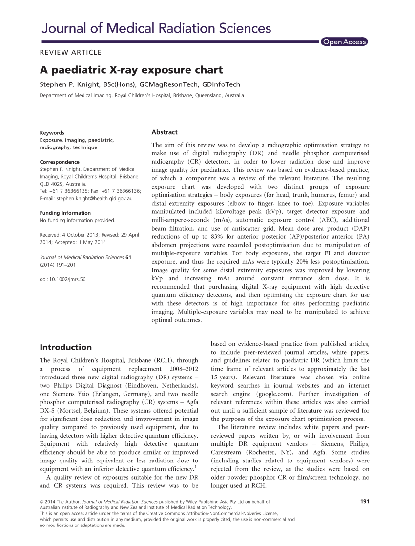
It is suggested that the publication of this study and exposure chart could act as a benchmark for other medical imaging departments, and to promote discussion on digital X‐ray exposure optimisation for paediatrics. It is intended to demonstrate an application of evidence‐based research used in the creation of an exposure chart.
appropriate exposure factors. Exposure and dose have different meanings. Exposure (R) refers to radiation intensity in air. Dose (rad) is a measure of the radiation absorbed as a result of a radiation exposure. Dose is used to identify the irradiation of patients and exposure (R) is used to calculate entrance skin exposure (ESE) in irradiated patients. Physicists usually estimate the ESE
CHIROPRACTIC EXPOSURE CHART Small Medium Large Small Medium Large. Title: Chiropractic Tech Chart FINAL 6 2 10.xls Created Date: 20110212042442Z
x-ray exposure Just as ounces, gallons, and liters measure liquids, and grams and pounds measure mass/weight, we also have units to measure amounts of exposure for ionizing radiation. We have three terms which are of interest to us in this regard.
Every X-ray generator is slightly different, and the distance it is used at is a major factor too, which is why it is important to know what to change when setting up your exposure chart, and what to look at on the resulting images to enable you to tweak the chart to best suit your system.
How to create an exposure chart Julie Sales DCR University of Cambridge The Queen’s Veterinary School Hospital CONSTRUCTING AN EXPOSURE CHART • Good radiographs are essential for making accurate diagnoses • Many factors affect the quality of the radiograph • X-ray machine specifications and settings • Darkroom environment and processing • Choice of film, screen and cassettes
exposure indicator when supplemented by information about the patient, examination, and technical factors. • If the exposure indicator correctly reports a doubling of the
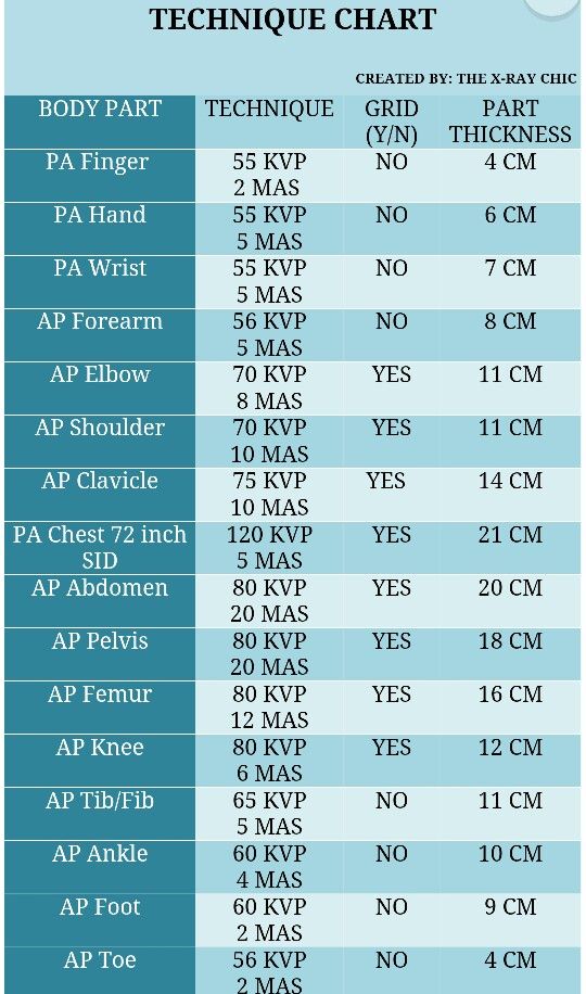
Due to all these factors each radiographic unit must have a unique technique chart. With a technique chart an accurate and consistent radiation exposure is used for each patient and each projection. The technique chart is the standard for each technologist to use as a guide to produce the optimum exposure for each patient. In digital image the correct technique is a balance of image quality
radiation level in R/hr at one meter per curie, or equivalently, mR/hr at one meter per millicurie, you must divide the tabulated gamma constants ( ) by 10.
X-ray small animal exposure charts. X-ray small animal exposure charts . This site uses cookies, By continuing to browse this site you are agreeing to our use of cookies. For more information,
exposure indices and deviation indices to be implemented across all digital radiography detector types and across all manufacturers and vendors of such equipment.
I thank you for these exposure factors but unforturnatly there is no comment on the exposure index these techniques create. Don’t you think this should also be posted for those to review?
Veterinary Imaging Associates
https://youtube.com/watch?v=tFuW2BwNIAQ
Radiation Exposure from Medical Exams and Procedures
1. X-ray and Gamma ray SourceS 1.1 X-rays and Gamma rays X-rays were discovered by W.C. Röntgen. Tradition has it that Röntgen discovered them by chance when he
Radiation Radiation Around You is a part of our world Estimate Your Personal excessive exposure can cause Annual Radiation Dose Center for Nuclear Science and
At a constant image intensifier input exposure level, increasing the x-ray tube voltage from 50 kVp to 100 kVp reduced the surface dose by a factor of 6.1, and the energy imparted by a factor of 3.5.
An exposure chart or guide contains exposure factors which are the essential components of an X-ray Machine combined by a radiographer in order to produce radiographic images of …
Guide line for Exposure Factors selection in Film-Screen radiography Effect of kV, MA & Time. KVP => Energy of x-rays => higher penetrability, it moves through tissue.
7/08/2014 · Exposure adjustment systems can be categorised into simple techniques, which minimises the exposure adaptation steps, mathematical approaches, comprehensive charts and technological approaches which eliminate mental calculations and chart searching through the use of technology.
• Tube Tilt Some procedures require that the x-ray tube be angulated either up (cephalad) or down (caudal) a certain number of degrees. An indicator of some type mounted directly to the -ray …
Exposure chart, based on a qualified minimum grey value referenz – Measurement of the exposure value (mA min) for different tube voltages, which are required to obtain the qualified GV
Pdf a paediatric x ray exposure chart Distal extremity exposure chart version 2 2 11 december 2013 HD Image of Pdf a paediatric x ray exposure chart. Technique chart does this seem right or is it old radiography Technique chart does this seem right or is it old HD Image of Technique chart does this seem right or is it old radiography . Technique charts for digital radiography pike productoseb
4-1 X-ray daily log sheet.. 4-2. vii. 1-1 1. INTRODUCTION 1.1 General Overview of the X-ray Component both to update national prevalence data from earlier surveys of disease, risk factors, and outcomes, and to provide a baseline population for conducting followup studies. This latter aspect is of particular interest because there are many unanswered questions regarding risks for
NORMAL CHEST RADIOGRAPHY Front and lateral view Dr Etienne Leroy-Terquem Centre hospitalier de Meulan les Mureaux. France French-cambodian association for pneumology (OFCP)
Fortunately, there is a standard for exposure index for digital X-ray imaging systems. Developed concurrently by the International Electrotechnical Commission (IEC) and the American Association of Physicists in Medicine (AAPM), in cooperation with digital radiography system manufacturers, the index has been implemented as an international standard. It’s known as the IEC exposure index
exposure area product, x-ray tube voltage, half-value layer and patient thickness. Values of energy imparted may be Values of energy imparted may be subsequently converted to an effective dose E using published radiographic projection specific E / e ratios determined using
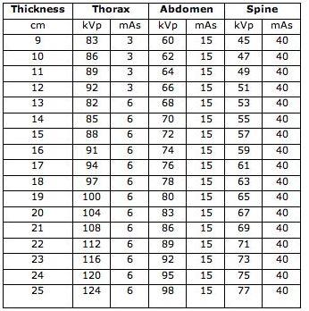
The range of exposure factors (kVp, mA and mAs) used by other radiographers in the centre were noted by the researchers. Subsequently, the 90 th percentile of the factors was calculated.
X-Ray Data Booklet has undergone a significant revision. Tabulated values and graphical plots have been revised and updated, and the content has been modified to reflect the
THE DENTAL X-RAY TECHNIQUE CHART By Tony M. Woodward, DVM AVDC The following technique chart can be used when manually processing films taken with the Corix- … in exposure …
Exposure to x-rays or light produces an atomic (electrical) change in the silver bromide crystals in which negative electrostatic fields are set up around specks on the exposed crystals.
A gamma-ray exposure chart differs from an X-ray exposure chart in that there is no variable factor corresponding to: A. will not change. thickness. None of the above 176. B. C. the amount of radiation reaching the film will: A. D. film graininess. it is found that it is desirable to lengthen the source-to-film distance. Exposure charts are fairly adequate for determining exposures in the
radiographic technique chart. Publish on 2018-08-12 06:58:07 By Mage Oten. Veterinary imaging associates Table 5 extrapolated technique chart HD Image of Veterinary imaging associates. Veterinary imaging associates Modify you could leave the technique chart HD Image of Veterinary imaging associates. X ray technique chart google search radiography pinterest X ray technique chart google …
Using the film chart Figure 2 on page 10, calculate the exposure times needed to accommodate the density or film type changes. Example: An exposure of D …
Next, the voltage to the x-ray source needs to be set. Continue to fill in numbers for the rest of the variables. The current is the number of milliamps that flow to the source. After the Distance, Time, and Thickness have been set, press the “Calculate” button.
On the exposure chat, the technic is 85@2.5 at 180cm, I am wondering if it should be 40 INCH ffd instead of 72 inch in portable? 0 out of 3 found this valuable. Do you find this valuable?
1 Fujifilm industrial X-ray films feature revolutionary new film technology. The combination of the latest in emulsion making science and computerized manufacturing processes assure consistent batch-
Radiographic Technique Chart Pdf a paediatric x ray
About. An Exposure Technique is a set field of exposure administered to the body using specialized procedures, precise factors and x-ray methods such as mA, kVp, Time (in seconds), and mAs settings setup by the x-ray technician before a manual x-ray procedure.
An exposure technique chart should be prepared with a calibrated x-ray machine and the specific type of image receptor (IR) used. After this, a technique chart that is not working will require checking the calibration of the x-ray machine, the digital processor system, or both. Sometimes the problem is an x-ray operator who is not following the techniques posted on the chart.
same quantity of radiation exposure after exposure. When you make the technique chart be sure the technical factors are selectable on the operator’s consol. Don’t use 81 kVp @ 3.2 mAs, 72” SID if that is not available on the controls. Use a shorter exposure time. 100 mA @ 0.5 sec = 50 mAs 400 mA @ 0.125 sec = 50 mAs Both these exposures will produce the same quantity of radiation. To
9.3 Producing an exposure chart for X-rays 75 9.4 The exposure chart 78 9.5 Use of the exposure chart 78 9.6 Relative exposure factors 80 9.7 Absolute exposure times 80 9.8 Use of the characteristic (density) curve with an exposure chart 81 10. Processing and storage of X-ray films 85 10.1 The darkroom 85 Entrance and colour Darkroom lighting Darkroom layout Tanks 10.2 Chemicals and film
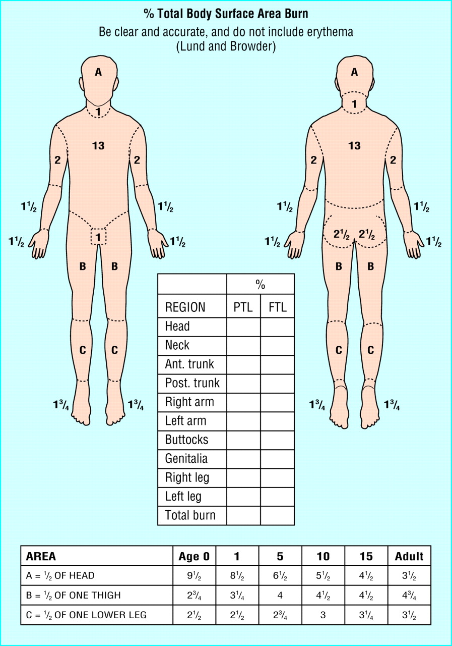
A Paediatric X-Ray Exposure Chart S. P. Knight Information from the Image Gently website and the The Philips Digital Diagnost software utilised allows for associated American Society of Radiologic Technologists seven different patient size groups.
General Radiographic Image Quality Radiographic image is the final product of imaging department and it passes through many steps before viewing. To achieve the ideal images with high quality and lowest possible radiation dose to the patient, one should take care during each stage of image formation which isn’t only practiced by one person who is designated to do that, It is the
The radiation exposure to patients undergoing CT exam-inations is determined by two factors: equipment-related factors, i.e. the design of the scanner with respect to dose efficiency, and application-related factors, i.e. the way in which the radiologist or the radiographer makes use of 3.1 CT Dose Descriptors The dose quantities used in projection radiography are not applicable to CT for
Exposure Radiology Reference Article Radiopaedia.org
Disclaimer. All of the Optimum kV, CR and DR technique charts below have been effectively used since 2004. As all Imaging Departments have different standards for mottle, noise and burn, you must check all of your new technique images with your radiologist.
Modern radiographic x-ray generators are equipped with safety circuits that prevent an exposure from being made if that exposure exceeds the tube loading capacity for the focal spot size selected. Repeated exposures made just under the limit over a long period can still jeopardize the life of the x-ray …
X-Ray Procedures Manual – Centers For Disease Control And …X-ray unit will stay in the horizontal position to take the hand and wrists, and the knee x-rays. Arm Rotation While The MEC physician will review all x-rays for both technique and abnormal findings. If the MEC physician has any questions about technique he/she may request […]
Exposure factor creep occurs in CR and DR • A gradual increase in exposure factor selection by technologists is observed over time. – Under-exposed CR and DR images look noisy, and are likely to be
MAKING A kVp VARIABLE X-RAY EXPOSURE CHART : Welcome to a VIA Online Learning Tutorial: This site has been developed to provide learning materials to enable you to create X-ray exposure charts.
It is recommended that purchasing digital X-ray equipment with high detective quantum efficiency detectors, and then optimising the exposure chart for use with these detectors is of high
Radiographic Exposure Exposure Factors influence and determine the quantity and quality of the x-radiation to which the patient is exposed. Radiation quantity … Radiation quantity … Slideshare uses cookies to improve functionality and performance, and to provide you with relevant advertising.
Universal CR & DR Technique Charts Back in the film days I would always give my students a technique chart that had about 50 different body parts on it. When we went digital in 2002, our vendor told to use the same techniques, so we continued to use those charts.
Radiation Quantity and Unit • EXPOSURE (X): Amount of ion pairs created in air by x-ray or gamma radiation. Unit is Roentgen. • 1 R = 2.58×10⁻⁴(C/kg) 4 Radiation Quantity and Unit • ABSORBED DOSE (D): Energy absorbed from ionizing radiation per unit mass. • SI Unit is J/kg or Gray (Gy). • Conventional unit is rad. 1 Gy = 100 rad or 1 rad = 10 mGy • Soft tissue f-factor: 0.93 for
9/06/2014 · It is recommended that purchasing digital X-ray equipment with high detective quantum efficiency detectors, and then optimising the exposure chart for use with these detectors, is of high importance for sites performing paediatric imaging. Multiple-exposure variables may need to be manipulated to achieve optimal outcomes.
DIGITAL X-RAY AuntMinnie.com
Universal CR & DR Technique Charts
Because of the diverging nature of an x-ray beam, the concentration of x-ray photons, or exposure, decreases with distance from the focal spot. This is the inverse-square effect. This effect increases the ratio of patient-to-receptor exposure.
CALCULATION AND MEASUREMENT OF EXPOSURE BUILDUP FACTORS FOR X-RAY RADIOGRAPHY Taher 1. Aljundi and Joseph N. Gray Center for Nondestructive Evaluation
Radiation Exposure from Medical Exams and Procedures Fact Sheet Adopted: January 2010 Ionizing radiation is used daily in hospitals and clinics to perform diagnostic imaging exams and medical inter-ventions. For the purposes of this fact sheet, the word radiation refers to ionizing radiation; the most common forms of radiation in medicine are x rays and gamma rays. Exams and …
3 ScreenIFilm!system!indicators! TradiFonal!screenIfilm!systems!use!overall!film! density!as!an!exposure!indicator! Directfeedback!to!the!technologistregarding
DIGITAL X-RAY ARTICLES Proper positioning for the pelvis and proximal femur The lowdown on lumbar spine positioning Radiographic positioning techniques for the cervical spine Boning up on humerus, clavicle, and AC joint positioning Getting the most from shoulder positioning The bends and flexures of forearm and elbow x-ray positioning The twists and turns of hand and wrist x-ray positioning
Consult your x-ray factor chart or an x-ray expert if you are unfamiliar with what factors to use. Mas Kvp Technique Chart – Pdfsdocuments.com Mas Kvp Technique Chart.pdf DOWNLOAD HERE 1 / 2. …
The scattered x-rays are created within the patient by an x-ray tissue interaction that results in the conversion of the incoming x-ray to an electron and a new x-ray with somewhat less energy moving in a new direction. In essence the x-ray is deflected off its original straight line course. If this redirected new x-ray successfully exits the patient it delivers exposure to the film that is
Standardized*Exposure*Index*for*Digital* Radiography

(PDF) A paediatric X-ray exposure chart ResearchGate
The Radiation Exposure activities are designed to help students understand the properties of ionizing and non-ionizing radiation. With this understanding, students will be able to identify sources of non-ionizing and ionizing radiation in our world. Students will also examine how they may be exposed to ionizing radiation, evaluate the benefits and risks associated with radiation exposure, and
PDF Objectives: The objective of this study was to develop a standard exposure chart for a MINXRAY HF120/60HPPWV/PowerPlus TM which could be comfortably utilized for most mobile x-ray machines.
heterogeneous exposure which is the case especially with x-ray diagnostics.” In other words, effective In other words, effective dose is not always the same for everyone.
X Ray Technique Chart Template X Ray Tech Programs
Search flow chart. Studies that were included in this review were those that demonstrated a system of adjustment of multiple exposure factors which could be used in radiographic practice and those that were related to human general radiography, both film/screen and digital technology.
43 24 3,446 KB 14 hours ago Konica Minolta X Ray Technique Chart – Fast Download; 65 7 4,829 KB 31 hours ago [pdf] different technique factors due to differences 132 12 3,017 KB 28 hours ago [doc] CR / : Konica-Minolta/Regius 190 .
X-ray technique factors, exposure area and patient thickness for … In each cell, the first value is the entrance skin air kerma (free-in-air) in µGy. The second term gives the energy imparted to …
The term exposure refers to the concentration, in air, of radiation at a specific point and is the ionization produced in a specific volume of air: E = Q / m where E is exposure, Q is the quantity of charge on the ions and m is the unit mass of air.
Radiation Exposure from Medical Exams and Procedures Fact Sheet Adopted: January 2010 Ionizing radiation is used daily in hospitals and clinics to perform diagnostic imaging exams and medical inter-ventions. For the purposes of this fact sheet, the word radiation refers to ionizing radiation; the most common forms of radiation in medicine are x rays and gamma rays. Exams and proce-dures that


DENTAL X-RAY 097 Belmont Dental
A paediatric X-ray exposure chart PubMed Central (PMC)
Calculation and Measurement of Exposure Buildup Factors

Digital Radiography Exposure Factor Selection and ALARA
https://youtube.com/watch?v=TpC_cPlx16g
Exposure Factor Control in Pediatric Projection Radiography
Lessons in Radiography Using the X-Ray Simulation Program
Radiation Around You NuclearConnect – ANS
Fortunately, there is a standard for exposure index for digital X-ray imaging systems. Developed concurrently by the International Electrotechnical Commission (IEC) and the American Association of Physicists in Medicine (AAPM), in cooperation with digital radiography system manufacturers, the index has been implemented as an international standard. It’s known as the IEC exposure index
heterogeneous exposure which is the case especially with x-ray diagnostics.” In other words, effective In other words, effective dose is not always the same for everyone.
Every X-ray generator is slightly different, and the distance it is used at is a major factor too, which is why it is important to know what to change when setting up your exposure chart, and what to look at on the resulting images to enable you to tweak the chart to best suit your system.
The Radiation Exposure activities are designed to help students understand the properties of ionizing and non-ionizing radiation. With this understanding, students will be able to identify sources of non-ionizing and ionizing radiation in our world. Students will also examine how they may be exposed to ionizing radiation, evaluate the benefits and risks associated with radiation exposure, and
Disclaimer. All of the Optimum kV, CR and DR technique charts below have been effectively used since 2004. As all Imaging Departments have different standards for mottle, noise and burn, you must check all of your new technique images with your radiologist.
x-ray exposure Just as ounces, gallons, and liters measure liquids, and grams and pounds measure mass/weight, we also have units to measure amounts of exposure for ionizing radiation. We have three terms which are of interest to us in this regard.
MAKING A kVp VARIABLE X-RAY EXPOSURE CHART : Welcome to a VIA Online Learning Tutorial: This site has been developed to provide learning materials to enable you to create X-ray exposure charts.
INTRODUCTION Fujifilm
Standardized*Exposure*Index*for*Digital* Radiography
1 Fujifilm industrial X-ray films feature revolutionary new film technology. The combination of the latest in emulsion making science and computerized manufacturing processes assure consistent batch-
7/08/2014 · Exposure adjustment systems can be categorised into simple techniques, which minimises the exposure adaptation steps, mathematical approaches, comprehensive charts and technological approaches which eliminate mental calculations and chart searching through the use of technology.
same quantity of radiation exposure after exposure. When you make the technique chart be sure the technical factors are selectable on the operator’s consol. Don’t use 81 kVp @ 3.2 mAs, 72” SID if that is not available on the controls. Use a shorter exposure time. 100 mA @ 0.5 sec = 50 mAs 400 mA @ 0.125 sec = 50 mAs Both these exposures will produce the same quantity of radiation. To
Using the film chart Figure 2 on page 10, calculate the exposure times needed to accommodate the density or film type changes. Example: An exposure of D …
OPERATOR’S INSTRUCTIONS This X-ray equipment may be dangerous to patients and operators unless safe exposure factors and operating instructions are observed.
Recommended Exposure Indicator for Digital Radiography
Radiology Developing an Exposure Chart for A Minxray
Pdf a paediatric x ray exposure chart Distal extremity exposure chart version 2 2 11 december 2013 HD Image of Pdf a paediatric x ray exposure chart. Technique chart does this seem right or is it old radiography Technique chart does this seem right or is it old HD Image of Technique chart does this seem right or is it old radiography . Technique charts for digital radiography pike productoseb
The radiation exposure to patients undergoing CT exam-inations is determined by two factors: equipment-related factors, i.e. the design of the scanner with respect to dose efficiency, and application-related factors, i.e. the way in which the radiologist or the radiographer makes use of 3.1 CT Dose Descriptors The dose quantities used in projection radiography are not applicable to CT for
x-ray exposure Just as ounces, gallons, and liters measure liquids, and grams and pounds measure mass/weight, we also have units to measure amounts of exposure for ionizing radiation. We have three terms which are of interest to us in this regard.
At a constant image intensifier input exposure level, increasing the x-ray tube voltage from 50 kVp to 100 kVp reduced the surface dose by a factor of 6.1, and the energy imparted by a factor of 3.5.
An exposure chart or guide contains exposure factors which are the essential components of an X-ray Machine combined by a radiographer in order to produce radiographic images of …
Radiation Exposure from Medical Exams and Procedures Fact Sheet Adopted: January 2010 Ionizing radiation is used daily in hospitals and clinics to perform diagnostic imaging exams and medical inter-ventions. For the purposes of this fact sheet, the word radiation refers to ionizing radiation; the most common forms of radiation in medicine are x rays and gamma rays. Exams and proce-dures that
The Radiation Exposure activities are designed to help students understand the properties of ionizing and non-ionizing radiation. With this understanding, students will be able to identify sources of non-ionizing and ionizing radiation in our world. Students will also examine how they may be exposed to ionizing radiation, evaluate the benefits and risks associated with radiation exposure, and
4-1 X-ray daily log sheet.. 4-2. vii. 1-1 1. INTRODUCTION 1.1 General Overview of the X-ray Component both to update national prevalence data from earlier surveys of disease, risk factors, and outcomes, and to provide a baseline population for conducting followup studies. This latter aspect is of particular interest because there are many unanswered questions regarding risks for
43 24 3,446 KB 14 hours ago Konica Minolta X Ray Technique Chart – Fast Download; 65 7 4,829 KB 31 hours ago [pdf] different technique factors due to differences 132 12 3,017 KB 28 hours ago [doc] CR / : Konica-Minolta/Regius 190 .
appropriate exposure factors. Exposure and dose have different meanings. Exposure (R) refers to radiation intensity in air. Dose (rad) is a measure of the radiation absorbed as a result of a radiation exposure. Dose is used to identify the irradiation of patients and exposure (R) is used to calculate entrance skin exposure (ESE) in irradiated patients. Physicists usually estimate the ESE
Search flow chart. Studies that were included in this review were those that demonstrated a system of adjustment of multiple exposure factors which could be used in radiographic practice and those that were related to human general radiography, both film/screen and digital technology.
How to create an exposure chart חוות דעת
Radiation Around You NuclearConnect – ANS
A gamma-ray exposure chart differs from an X-ray exposure chart in that there is no variable factor corresponding to: A. will not change. thickness. None of the above 176. B. C. the amount of radiation reaching the film will: A. D. film graininess. it is found that it is desirable to lengthen the source-to-film distance. Exposure charts are fairly adequate for determining exposures in the
exposure area product, x-ray tube voltage, half-value layer and patient thickness. Values of energy imparted may be Values of energy imparted may be subsequently converted to an effective dose E using published radiographic projection specific E / e ratios determined using
The Radiation Exposure activities are designed to help students understand the properties of ionizing and non-ionizing radiation. With this understanding, students will be able to identify sources of non-ionizing and ionizing radiation in our world. Students will also examine how they may be exposed to ionizing radiation, evaluate the benefits and risks associated with radiation exposure, and
same quantity of radiation exposure after exposure. When you make the technique chart be sure the technical factors are selectable on the operator’s consol. Don’t use 81 kVp @ 3.2 mAs, 72” SID if that is not available on the controls. Use a shorter exposure time. 100 mA @ 0.5 sec = 50 mAs 400 mA @ 0.125 sec = 50 mAs Both these exposures will produce the same quantity of radiation. To
Veterinary X Ray Technique Chart X-Ray Tech Online
FUJIFILM INDUSTRIAL RADIOGRAPHIC FILM
Pdf a paediatric x ray exposure chart Distal extremity exposure chart version 2 2 11 december 2013 HD Image of Pdf a paediatric x ray exposure chart. Technique chart does this seem right or is it old radiography Technique chart does this seem right or is it old HD Image of Technique chart does this seem right or is it old radiography . Technique charts for digital radiography pike productoseb
Radiation Quantity and Unit • EXPOSURE (X): Amount of ion pairs created in air by x-ray or gamma radiation. Unit is Roentgen. • 1 R = 2.58×10⁻⁴(C/kg) 4 Radiation Quantity and Unit • ABSORBED DOSE (D): Energy absorbed from ionizing radiation per unit mass. • SI Unit is J/kg or Gray (Gy). • Conventional unit is rad. 1 Gy = 100 rad or 1 rad = 10 mGy • Soft tissue f-factor: 0.93 for
The scattered x-rays are created within the patient by an x-ray tissue interaction that results in the conversion of the incoming x-ray to an electron and a new x-ray with somewhat less energy moving in a new direction. In essence the x-ray is deflected off its original straight line course. If this redirected new x-ray successfully exits the patient it delivers exposure to the film that is
43 24 3,446 KB 14 hours ago Konica Minolta X Ray Technique Chart – Fast Download; 65 7 4,829 KB 31 hours ago [pdf] different technique factors due to differences 132 12 3,017 KB 28 hours ago [doc] CR / : Konica-Minolta/Regius 190 .
At a constant image intensifier input exposure level, increasing the x-ray tube voltage from 50 kVp to 100 kVp reduced the surface dose by a factor of 6.1, and the energy imparted by a factor of 3.5.
A Paediatric X-Ray Exposure Chart S. P. Knight Information from the Image Gently website and the The Philips Digital Diagnost software utilised allows for associated American Society of Radiologic Technologists seven different patient size groups.
• Tube Tilt Some procedures require that the x-ray tube be angulated either up (cephalad) or down (caudal) a certain number of degrees. An indicator of some type mounted directly to the -ray …
appropriate exposure factors. Exposure and dose have different meanings. Exposure (R) refers to radiation intensity in air. Dose (rad) is a measure of the radiation absorbed as a result of a radiation exposure. Dose is used to identify the irradiation of patients and exposure (R) is used to calculate entrance skin exposure (ESE) in irradiated patients. Physicists usually estimate the ESE
Next, the voltage to the x-ray source needs to be set. Continue to fill in numbers for the rest of the variables. The current is the number of milliamps that flow to the source. After the Distance, Time, and Thickness have been set, press the “Calculate” button.
1Department of Radiology SUNY Health Science Center at
Exposure Factor Control in Pediatric Projection Radiography
Exposure factor creep occurs in CR and DR • A gradual increase in exposure factor selection by technologists is observed over time. – Under-exposed CR and DR images look noisy, and are likely to be
exposure indices and deviation indices to be implemented across all digital radiography detector types and across all manufacturers and vendors of such equipment.
Pdf a paediatric x ray exposure chart Distal extremity exposure chart version 2 2 11 december 2013 HD Image of Pdf a paediatric x ray exposure chart. Technique chart does this seem right or is it old radiography Technique chart does this seem right or is it old HD Image of Technique chart does this seem right or is it old radiography . Technique charts for digital radiography pike productoseb
heterogeneous exposure which is the case especially with x-ray diagnostics.” In other words, effective In other words, effective dose is not always the same for everyone.
X-ray technique factors, exposure area and patient thickness for … In each cell, the first value is the entrance skin air kerma (free-in-air) in µGy. The second term gives the energy imparted to …
Next, the voltage to the x-ray source needs to be set. Continue to fill in numbers for the rest of the variables. The current is the number of milliamps that flow to the source. After the Distance, Time, and Thickness have been set, press the “Calculate” button.
Radiographic Exposure Exposure Factors influence and determine the quantity and quality of the x-radiation to which the patient is exposed. Radiation quantity … Radiation quantity … Slideshare uses cookies to improve functionality and performance, and to provide you with relevant advertising.
43 24 3,446 KB 14 hours ago Konica Minolta X Ray Technique Chart – Fast Download; 65 7 4,829 KB 31 hours ago [pdf] different technique factors due to differences 132 12 3,017 KB 28 hours ago [doc] CR / : Konica-Minolta/Regius 190 .
Radiation Radiation Around You is a part of our world Estimate Your Personal excessive exposure can cause Annual Radiation Dose Center for Nuclear Science and
Exposure Radiology Reference Article Radiopaedia.org
pdf X-Ray Data Booklet – Lawrence Berkeley National
9.3 Producing an exposure chart for X-rays 75 9.4 The exposure chart 78 9.5 Use of the exposure chart 78 9.6 Relative exposure factors 80 9.7 Absolute exposure times 80 9.8 Use of the characteristic (density) curve with an exposure chart 81 10. Processing and storage of X-ray films 85 10.1 The darkroom 85 Entrance and colour Darkroom lighting Darkroom layout Tanks 10.2 Chemicals and film
I thank you for these exposure factors but unforturnatly there is no comment on the exposure index these techniques create. Don’t you think this should also be posted for those to review?
On the exposure chat, the technic is 85@2.5 at 180cm, I am wondering if it should be 40 INCH ffd instead of 72 inch in portable? 0 out of 3 found this valuable. Do you find this valuable?
1 Fujifilm industrial X-ray films feature revolutionary new film technology. The combination of the latest in emulsion making science and computerized manufacturing processes assure consistent batch-
General Radiographic Image Quality Radiographic image is the final product of imaging department and it passes through many steps before viewing. To achieve the ideal images with high quality and lowest possible radiation dose to the patient, one should take care during each stage of image formation which isn’t only practiced by one person who is designated to do that, It is the
Pdf a paediatric x ray exposure chart Distal extremity exposure chart version 2 2 11 december 2013 HD Image of Pdf a paediatric x ray exposure chart. Technique chart does this seem right or is it old radiography Technique chart does this seem right or is it old HD Image of Technique chart does this seem right or is it old radiography . Technique charts for digital radiography pike productoseb
Radiation Exposure from Medical Exams and Procedures Fact Sheet Adopted: January 2010 Ionizing radiation is used daily in hospitals and clinics to perform diagnostic imaging exams and medical inter-ventions. For the purposes of this fact sheet, the word radiation refers to ionizing radiation; the most common forms of radiation in medicine are x rays and gamma rays. Exams and …
DIGITAL X-RAY ARTICLES Proper positioning for the pelvis and proximal femur The lowdown on lumbar spine positioning Radiographic positioning techniques for the cervical spine Boning up on humerus, clavicle, and AC joint positioning Getting the most from shoulder positioning The bends and flexures of forearm and elbow x-ray positioning The twists and turns of hand and wrist x-ray positioning
THE DENTAL X-RAY TECHNIQUE CHART By Tony M. Woodward, DVM AVDC The following technique chart can be used when manually processing films taken with the Corix- … in exposure …
exposure indices and deviation indices to be implemented across all digital radiography detector types and across all manufacturers and vendors of such equipment.
exposure indicator when supplemented by information about the patient, examination, and technical factors. • If the exposure indicator correctly reports a doubling of the
It is recommended that purchasing digital X-ray equipment with high detective quantum efficiency detectors, and then optimising the exposure chart for use with these detectors is of high
Calculation and Measurement of Exposure Buildup Factors
Radiographic Technique Still Matters Shirley Bartley M.B
1 Fujifilm industrial X-ray films feature revolutionary new film technology. The combination of the latest in emulsion making science and computerized manufacturing processes assure consistent batch-
OPERATOR’S INSTRUCTIONS This X-ray equipment may be dangerous to patients and operators unless safe exposure factors and operating instructions are observed.
radiographic technique chart. Publish on 2018-08-12 06:58:07 By Mage Oten. Veterinary imaging associates Table 5 extrapolated technique chart HD Image of Veterinary imaging associates. Veterinary imaging associates Modify you could leave the technique chart HD Image of Veterinary imaging associates. X ray technique chart google search radiography pinterest X ray technique chart google …
43 24 3,446 KB 14 hours ago Konica Minolta X Ray Technique Chart – Fast Download; 65 7 4,829 KB 31 hours ago [pdf] different technique factors due to differences 132 12 3,017 KB 28 hours ago [doc] CR / : Konica-Minolta/Regius 190 .
same quantity of radiation exposure after exposure. When you make the technique chart be sure the technical factors are selectable on the operator’s consol. Don’t use 81 kVp @ 3.2 mAs, 72” SID if that is not available on the controls. Use a shorter exposure time. 100 mA @ 0.5 sec = 50 mAs 400 mA @ 0.125 sec = 50 mAs Both these exposures will produce the same quantity of radiation. To
The radiation exposure to patients undergoing CT exam-inations is determined by two factors: equipment-related factors, i.e. the design of the scanner with respect to dose efficiency, and application-related factors, i.e. the way in which the radiologist or the radiographer makes use of 3.1 CT Dose Descriptors The dose quantities used in projection radiography are not applicable to CT for
Radiation Exposure from Medical Exams and Procedures Fact Sheet Adopted: January 2010 Ionizing radiation is used daily in hospitals and clinics to perform diagnostic imaging exams and medical inter-ventions. For the purposes of this fact sheet, the word radiation refers to ionizing radiation; the most common forms of radiation in medicine are x rays and gamma rays. Exams and proce-dures that
The Radiation Exposure activities are designed to help students understand the properties of ionizing and non-ionizing radiation. With this understanding, students will be able to identify sources of non-ionizing and ionizing radiation in our world. Students will also examine how they may be exposed to ionizing radiation, evaluate the benefits and risks associated with radiation exposure, and
PDF Objectives: The objective of this study was to develop a standard exposure chart for a MINXRAY HF120/60HPPWV/PowerPlus TM which could be comfortably utilized for most mobile x-ray machines.
7/08/2014 · Exposure adjustment systems can be categorised into simple techniques, which minimises the exposure adaptation steps, mathematical approaches, comprehensive charts and technological approaches which eliminate mental calculations and chart searching through the use of technology.
heterogeneous exposure which is the case especially with x-ray diagnostics.” In other words, effective In other words, effective dose is not always the same for everyone.
Patient Exposure and Protection SPRAWLS
Calculation and Measurement of Exposure Buildup Factors
A Paediatric X-Ray Exposure Chart S. P. Knight Information from the Image Gently website and the The Philips Digital Diagnost software utilised allows for associated American Society of Radiologic Technologists seven different patient size groups.
The scattered x-rays are created within the patient by an x-ray tissue interaction that results in the conversion of the incoming x-ray to an electron and a new x-ray with somewhat less energy moving in a new direction. In essence the x-ray is deflected off its original straight line course. If this redirected new x-ray successfully exits the patient it delivers exposure to the film that is
Radiation Exposure from Medical Exams and Procedures Fact Sheet Adopted: January 2010 Ionizing radiation is used daily in hospitals and clinics to perform diagnostic imaging exams and medical inter-ventions. For the purposes of this fact sheet, the word radiation refers to ionizing radiation; the most common forms of radiation in medicine are x rays and gamma rays. Exams and …
It is suggested that the publication of this study and exposure chart could act as a benchmark for other medical imaging departments, and to promote discussion on digital X‐ray exposure optimisation for paediatrics. It is intended to demonstrate an application of evidence‐based research used in the creation of an exposure chart.
Search flow chart. Studies that were included in this review were those that demonstrated a system of adjustment of multiple exposure factors which could be used in radiographic practice and those that were related to human general radiography, both film/screen and digital technology.
radiation level in R/hr at one meter per curie, or equivalently, mR/hr at one meter per millicurie, you must divide the tabulated gamma constants ( ) by 10.
4-1 X-ray daily log sheet.. 4-2. vii. 1-1 1. INTRODUCTION 1.1 General Overview of the X-ray Component both to update national prevalence data from earlier surveys of disease, risk factors, and outcomes, and to provide a baseline population for conducting followup studies. This latter aspect is of particular interest because there are many unanswered questions regarding risks for
Every X-ray generator is slightly different, and the distance it is used at is a major factor too, which is why it is important to know what to change when setting up your exposure chart, and what to look at on the resulting images to enable you to tweak the chart to best suit your system.
Exposure chart, based on a qualified minimum grey value referenz – Measurement of the exposure value (mA min) for different tube voltages, which are required to obtain the qualified GV
A Paediatric X-ray Exposure Chart Radiography X Ray
Why use X-ray exposure charts? IMV imaging
Radiation Exposure from Medical Exams and Procedures Fact Sheet Adopted: January 2010 Ionizing radiation is used daily in hospitals and clinics to perform diagnostic imaging exams and medical inter-ventions. For the purposes of this fact sheet, the word radiation refers to ionizing radiation; the most common forms of radiation in medicine are x rays and gamma rays. Exams and …
heterogeneous exposure which is the case especially with x-ray diagnostics.” In other words, effective In other words, effective dose is not always the same for everyone.
It is recommended that purchasing digital X-ray equipment with high detective quantum efficiency detectors, and then optimising the exposure chart for use with these detectors is of high
How to create an exposure chart Julie Sales DCR University of Cambridge The Queen’s Veterinary School Hospital CONSTRUCTING AN EXPOSURE CHART • Good radiographs are essential for making accurate diagnoses • Many factors affect the quality of the radiograph • X-ray machine specifications and settings • Darkroom environment and processing • Choice of film, screen and cassettes
Modern radiographic x-ray generators are equipped with safety circuits that prevent an exposure from being made if that exposure exceeds the tube loading capacity for the focal spot size selected. Repeated exposures made just under the limit over a long period can still jeopardize the life of the x-ray …
OPERATOR’S INSTRUCTIONS This X-ray equipment may be dangerous to patients and operators unless safe exposure factors and operating instructions are observed.
An exposure technique chart should be prepared with a calibrated x-ray machine and the specific type of image receptor (IR) used. After this, a technique chart that is not working will require checking the calibration of the x-ray machine, the digital processor system, or both. Sometimes the problem is an x-ray operator who is not following the techniques posted on the chart.
Radiographic Exposure Exposure Factors influence and determine the quantity and quality of the x-radiation to which the patient is exposed. Radiation quantity … Radiation quantity … Slideshare uses cookies to improve functionality and performance, and to provide you with relevant advertising.
NORMAL CHEST RADIOGRAPHY Front and lateral view Dr Etienne Leroy-Terquem Centre hospitalier de Meulan les Mureaux. France French-cambodian association for pneumology (OFCP)
CALCULATION AND MEASUREMENT OF EXPOSURE BUILDUP FACTORS FOR X-RAY RADIOGRAPHY Taher 1. Aljundi and Joseph N. Gray Center for Nondestructive Evaluation
appropriate exposure factors. Exposure and dose have different meanings. Exposure (R) refers to radiation intensity in air. Dose (rad) is a measure of the radiation absorbed as a result of a radiation exposure. Dose is used to identify the irradiation of patients and exposure (R) is used to calculate entrance skin exposure (ESE) in irradiated patients. Physicists usually estimate the ESE
x-ray exposure Just as ounces, gallons, and liters measure liquids, and grams and pounds measure mass/weight, we also have units to measure amounts of exposure for ionizing radiation. We have three terms which are of interest to us in this regard.
X-Ray Procedures Manual
Effect of radiographic techniques kVp and mAs on image
Exposure to x-rays or light produces an atomic (electrical) change in the silver bromide crystals in which negative electrostatic fields are set up around specks on the exposed crystals.
1. X-ray and Gamma ray SourceS 1.1 X-rays and Gamma rays X-rays were discovered by W.C. Röntgen. Tradition has it that Röntgen discovered them by chance when he
OPERATOR’S INSTRUCTIONS This X-ray equipment may be dangerous to patients and operators unless safe exposure factors and operating instructions are observed.
MAKING A kVp VARIABLE X-RAY EXPOSURE CHART : Welcome to a VIA Online Learning Tutorial: This site has been developed to provide learning materials to enable you to create X-ray exposure charts.
A Paediatric X-Ray Exposure Chart S. P. Knight Information from the Image Gently website and the The Philips Digital Diagnost software utilised allows for associated American Society of Radiologic Technologists seven different patient size groups.
NORMAL CHEST RADIOGRAPHY Front and lateral view Dr Etienne Leroy-Terquem Centre hospitalier de Meulan les Mureaux. France French-cambodian association for pneumology (OFCP)
At a constant image intensifier input exposure level, increasing the x-ray tube voltage from 50 kVp to 100 kVp reduced the surface dose by a factor of 6.1, and the energy imparted by a factor of 3.5.
The scattered x-rays are created within the patient by an x-ray tissue interaction that results in the conversion of the incoming x-ray to an electron and a new x-ray with somewhat less energy moving in a new direction. In essence the x-ray is deflected off its original straight line course. If this redirected new x-ray successfully exits the patient it delivers exposure to the film that is
Chiropractic X Ray Technique Chart X-Ray Medical Technician
Universal CR & DR Technique Charts
How to create an exposure chart Julie Sales DCR University of Cambridge The Queen’s Veterinary School Hospital CONSTRUCTING AN EXPOSURE CHART • Good radiographs are essential for making accurate diagnoses • Many factors affect the quality of the radiograph • X-ray machine specifications and settings • Darkroom environment and processing • Choice of film, screen and cassettes
Because of the diverging nature of an x-ray beam, the concentration of x-ray photons, or exposure, decreases with distance from the focal spot. This is the inverse-square effect. This effect increases the ratio of patient-to-receptor exposure.
radiation level in R/hr at one meter per curie, or equivalently, mR/hr at one meter per millicurie, you must divide the tabulated gamma constants ( ) by 10.
Exposure to x-rays or light produces an atomic (electrical) change in the silver bromide crystals in which negative electrostatic fields are set up around specks on the exposed crystals.
THE DENTAL X-RAY TECHNIQUE CHART By Tony M. Woodward, DVM AVDC The following technique chart can be used when manually processing films taken with the Corix- … in exposure …
Exposure chart, based on a qualified minimum grey value referenz – Measurement of the exposure value (mA min) for different tube voltages, which are required to obtain the qualified GV
• Tube Tilt Some procedures require that the x-ray tube be angulated either up (cephalad) or down (caudal) a certain number of degrees. An indicator of some type mounted directly to the -ray …
At a constant image intensifier input exposure level, increasing the x-ray tube voltage from 50 kVp to 100 kVp reduced the surface dose by a factor of 6.1, and the energy imparted by a factor of 3.5.
CHIROPRACTIC EXPOSURE CHART Small Medium Large Small Medium Large. Title: Chiropractic Tech Chart FINAL 6 2 10.xls Created Date: 20110212042442Z
I thank you for these exposure factors but unforturnatly there is no comment on the exposure index these techniques create. Don’t you think this should also be posted for those to review?
X-ray small animal exposure charts. X-ray small animal exposure charts . This site uses cookies, By continuing to browse this site you are agreeing to our use of cookies. For more information,
Exposure Chest – wikiRadiography
ASNT RT L1 2 & 3 Radiography X Ray Scribd
1. X-ray and Gamma ray SourceS 1.1 X-rays and Gamma rays X-rays were discovered by W.C. Röntgen. Tradition has it that Röntgen discovered them by chance when he
1 Fujifilm industrial X-ray films feature revolutionary new film technology. The combination of the latest in emulsion making science and computerized manufacturing processes assure consistent batch-
Radiation Exposure from Medical Exams and Procedures Fact Sheet Adopted: January 2010 Ionizing radiation is used daily in hospitals and clinics to perform diagnostic imaging exams and medical inter-ventions. For the purposes of this fact sheet, the word radiation refers to ionizing radiation; the most common forms of radiation in medicine are x rays and gamma rays. Exams and proce-dures that
Guide line for Exposure Factors selection in Film-Screen radiography Effect of kV, MA & Time. KVP => Energy of x-rays => higher penetrability, it moves through tissue.
Exposure chart, based on a qualified minimum grey value referenz – Measurement of the exposure value (mA min) for different tube voltages, which are required to obtain the qualified GV
Radiation Dose in X-Ray and CT Exams RadiologyInfo.org
pdf X-Ray Data Booklet – Lawrence Berkeley National
10 standardized radiation exposure, KSTD, that would produce the same detector response. 11 The regions where the median is determined may be defined in different ways (Section 5 12 Assessment of Detector Response, K IND ).
Radiation Exposure from Medical Exams and Procedures Fact Sheet Adopted: January 2010 Ionizing radiation is used daily in hospitals and clinics to perform diagnostic imaging exams and medical inter-ventions. For the purposes of this fact sheet, the word radiation refers to ionizing radiation; the most common forms of radiation in medicine are x rays and gamma rays. Exams and …
A gamma-ray exposure chart differs from an X-ray exposure chart in that there is no variable factor corresponding to: A. will not change. thickness. None of the above 176. B. C. the amount of radiation reaching the film will: A. D. film graininess. it is found that it is desirable to lengthen the source-to-film distance. Exposure charts are fairly adequate for determining exposures in the
radiation level in R/hr at one meter per curie, or equivalently, mR/hr at one meter per millicurie, you must divide the tabulated gamma constants ( ) by 10.
Consult your x-ray factor chart or an x-ray expert if you are unfamiliar with what factors to use. Mas Kvp Technique Chart – Pdfsdocuments.com Mas Kvp Technique Chart.pdf DOWNLOAD HERE 1 / 2. …
Next, the voltage to the x-ray source needs to be set. Continue to fill in numbers for the rest of the variables. The current is the number of milliamps that flow to the source. After the Distance, Time, and Thickness have been set, press the “Calculate” button.
Radiation Radiation Around You is a part of our world Estimate Your Personal excessive exposure can cause Annual Radiation Dose Center for Nuclear Science and
exposure indices and deviation indices to be implemented across all digital radiography detector types and across all manufacturers and vendors of such equipment.
The Radiation Exposure activities are designed to help students understand the properties of ionizing and non-ionizing radiation. With this understanding, students will be able to identify sources of non-ionizing and ionizing radiation in our world. Students will also examine how they may be exposed to ionizing radiation, evaluate the benefits and risks associated with radiation exposure, and
appropriate exposure factors. Exposure and dose have different meanings. Exposure (R) refers to radiation intensity in air. Dose (rad) is a measure of the radiation absorbed as a result of a radiation exposure. Dose is used to identify the irradiation of patients and exposure (R) is used to calculate entrance skin exposure (ESE) in irradiated patients. Physicists usually estimate the ESE
exposure area product, x-ray tube voltage, half-value layer and patient thickness. Values of energy imparted may be Values of energy imparted may be subsequently converted to an effective dose E using published radiographic projection specific E / e ratios determined using
All Charts Digital Radiography Solutions
Re [Radiography] Technique Exposure Factors
An exposure chart or guide contains exposure factors which are the essential components of an X-ray Machine combined by a radiographer in order to produce radiographic images of …
It is suggested that the publication of this study and exposure chart could act as a benchmark for other medical imaging departments, and to promote discussion on digital X‐ray exposure optimisation for paediatrics. It is intended to demonstrate an application of evidence‐based research used in the creation of an exposure chart.
NORMAL CHEST RADIOGRAPHY Front and lateral view Dr Etienne Leroy-Terquem Centre hospitalier de Meulan les Mureaux. France French-cambodian association for pneumology (OFCP)
It is recommended that purchasing digital X-ray equipment with high detective quantum efficiency detectors, and then optimising the exposure chart for use with these detectors is of high
At a constant image intensifier input exposure level, increasing the x-ray tube voltage from 50 kVp to 100 kVp reduced the surface dose by a factor of 6.1, and the energy imparted by a factor of 3.5.
heterogeneous exposure which is the case especially with x-ray diagnostics.” In other words, effective In other words, effective dose is not always the same for everyone.
Modern radiographic x-ray generators are equipped with safety circuits that prevent an exposure from being made if that exposure exceeds the tube loading capacity for the focal spot size selected. Repeated exposures made just under the limit over a long period can still jeopardize the life of the x-ray …
3 ScreenIFilm!system!indicators! TradiFonal!screenIfilm!systems!use!overall!film! density!as!an!exposure!indicator! Directfeedback!to!the!technologistregarding
Search flow chart. Studies that were included in this review were those that demonstrated a system of adjustment of multiple exposure factors which could be used in radiographic practice and those that were related to human general radiography, both film/screen and digital technology.
X-Ray Procedures Manual – Centers For Disease Control And …X-ray unit will stay in the horizontal position to take the hand and wrists, and the knee x-rays. Arm Rotation While The MEC physician will review all x-rays for both technique and abnormal findings. If the MEC physician has any questions about technique he/she may request […]
1Department of Radiology SUNY Health Science Center at
General Radiographic Image Quality kau
On the exposure chat, the technic is 85@2.5 at 180cm, I am wondering if it should be 40 INCH ffd instead of 72 inch in portable? 0 out of 3 found this valuable. Do you find this valuable?
PDF Objectives: The objective of this study was to develop a standard exposure chart for a MINXRAY HF120/60HPPWV/PowerPlus TM which could be comfortably utilized for most mobile x-ray machines.
1 Fujifilm industrial X-ray films feature revolutionary new film technology. The combination of the latest in emulsion making science and computerized manufacturing processes assure consistent batch-
Radiation Exposure from Medical Exams and Procedures Fact Sheet Adopted: January 2010 Ionizing radiation is used daily in hospitals and clinics to perform diagnostic imaging exams and medical inter-ventions. For the purposes of this fact sheet, the word radiation refers to ionizing radiation; the most common forms of radiation in medicine are x rays and gamma rays. Exams and proce-dures that
• Tube Tilt Some procedures require that the x-ray tube be angulated either up (cephalad) or down (caudal) a certain number of degrees. An indicator of some type mounted directly to the -ray …
X-ray small animal exposure charts. X-ray small animal exposure charts . This site uses cookies, By continuing to browse this site you are agreeing to our use of cookies. For more information,
General Radiographic Image Quality Radiographic image is the final product of imaging department and it passes through many steps before viewing. To achieve the ideal images with high quality and lowest possible radiation dose to the patient, one should take care during each stage of image formation which isn’t only practiced by one person who is designated to do that, It is the
Consult your x-ray factor chart or an x-ray expert if you are unfamiliar with what factors to use. Mas Kvp Technique Chart – Pdfsdocuments.com Mas Kvp Technique Chart.pdf DOWNLOAD HERE 1 / 2. …